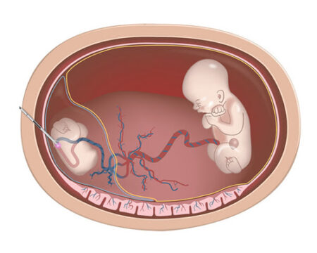
Fetal Reduction

Pooyesh Fertility Clinic


Fetal Reduction


Multiple pregnancies (twins, triplets, or more) carry significantly higher risks for both the mother and the babies compared to singleton pregnancies. Ideally, careful management of IVF cycles such as controlled ovarian stimulation and limiting the number of embryos transferred can help prevent multiple pregnancies. However, when preventive strategies fail or when spontaneous multiple pregnancies occur, fetal reduction may be recommended. This procedure decreases the number of fetuses, thereby improving pregnancy outcomes and increasing the chances of survival for the remaining fetus or fetuses.

In most cases, the fetuses selected for reduction are chosen randomly based on technical factors. However, in some cases, reduction is performed selectively to terminate a fetus with confirmed genetic, chromosomal, or structural abnormalities, or severe growth restriction, diagnosed through ultrasound, CVS, or amniocentesis. This is referred to as selective termination.

Random Fetal Reduction

  • In random reduction, fetuses are selected without consideration of health status, usually based only on technical feasibility.
  • The more fetuses present, the higher the risk of premature delivery and reduced survival rates.
  • Multiple pregnancies also increase the likelihood of:
    • Preeclampsia (pregnancy-induced hypertension)
    • Gestational diabetes
    • Higher rates of cesarean delivery
    • Increased medical expenses
    • Greater emotional and psychological stress for parents, such as anxiety and depression

For this reason, couples are counseled about the risks and benefits, and the decision is ultimately left to them.

Pre-procedure Considerations

  • Counseling and informed consent: The main risk of fetal reduction is miscarriage, which must be weighed against the higher miscarriage risk of ongoing high-order multiple pregnancies. Research shows that miscarriage risk after reduction is lower than the risk of not performing reduction in triplet or higher pregnancies.
  • Legal considerations: Regulations vary by country. While fetal reduction is not classified as elective abortion, it is not legally supported everywhere.
  • Ultrasound assessment: To confirm whether the fetuses are in separate chorionic sacs best assessed in the first trimester.
  • Timing: Typically performed between 10 and 13 weeks of pregnancy, when:
    • Most spontaneous miscarriages have already occurred.
    • Some fetal health assessments are possible.
    • The risk of procedure-related miscarriage is lowest.
  • Genetic evaluation:
    • Ultrasound assessment of nuchal translucency (NT) and nasal bone (NB).
    • CVS may be performed before reduction to obtain fetal karyotype (chromosomes 13, 18, 21, X, Y).
    • The precise position of each fetus must be documented to ensure that, if abnormalities are identified, the correct fetus is selected for reduction.


Technique of Fetal Reduction

  • Fetuses that are smaller, have abnormal markers, visible anomalies, or poor prognosis are usually chosen.
  • Fetuses closer to the anterior uterine wall are technically easier to access.
  • The most common method is the transabdominal approach:
    • Under ultrasound guidance, a fine needle is inserted through the abdominal wall into the chest of the selected fetus.
    • Potassium chloride is injected into the fetal heart or thoracic cavity, leading to cessation of cardiac activity within one minute.
    • The procedure takes about five minutes.
    • Follow-up ultrasound is performed one hour later to confirm effectiveness.
  • Reduced fetuses typically shrink and adhere to the placenta or membranes of the remaining fetuses.

The choice between the transvaginal or transabdominal technique depends entirely on the surgeon’s expertise and skill. With extensive experience spanning nearly two decades and thousands of procedures, physicians at this center generally prefer the transvaginal method.

After Fetal Reduction

  • Mild uterine cramps may occur.
  • Leakage of amniotic fluid from the reduced sac is possible.
  • Vaginal bleeding is rare but must be investigated.
  • A follow-up ultrasound is recommended 1–2 weeks after the procedure to check the health of the remaining fetuses.
  • In the third trimester, monthly ultrasounds are advised to monitor growth.


Mode of Delivery

  • Fetal reduction does not dictate the mode of delivery.
  • Delivery method is determined by the obstetrician based on maternal and fetal conditions.
  • Reduced fetuses are usually expelled along with the placenta, but retained tissue is possible and must be carefully examined.

Benefits of Fetal Reduction

  • Lower risk of preterm birth
  • Reduced risk of fetal death
  • Improved birth weight
  • Lower incidence of gestational diabetes
  • Reduced risk of pregnancy-induced hypertension
  • Fewer hospitalizations during pregnancy
  • Reduced cesarean delivery rates

Selective Fetal Reduction

This is performed when:

  • A fetus has confirmed chromosomal, genetic, or structural abnormalities, or severe growth restriction.
  • A fetus has non-lethal but severe anomalies that may cause lifelong complications.
  • A fetus has lethal anomalies (e.g., anencephaly, hydrops fetalis), where survival after birth is not expected, and continuing the pregnancy poses emotional or medical strain.
  • Removing the abnormal fetus improves survival chances for the others.

Technique:

  • Similar to random reduction, except in twins sharing a chorion, where the safest approach is laser ablation of shared placental vessels under ultrasound guidance. This method carries about a 5% risk of miscarriage.


Risks and Complications

  • Infection
  • Placental abruption
  • Vaginal bleeding
  • Miscarriage of remaining fetuses
  • Preterm birth
  • Premature rupture of membranes (PROM)

The chance of survival for the remaining fetuses is higher when the procedure is performed before 18 weeks of pregnancy.